In this tutorial we will see examples of how to manipulate images in DICOM files with the pydicom and ImageIO libraries.
Basic documentation on reading, accessing, modifying and writing DICOM files
pydicom
imageio
Libraries and packages
!pip install pydicom
Collecting pydicom
[?25l Downloading https://files.pythonhosted.org/packages/f4/15/df16546bc59bfca390cf072d473fb2c8acd4231636f64356593a63137e55/pydicom-2.1.2-py3-none-any.whl (1.9MB)
[K |████████████████████████████████| 1.9MB 5.4MB/s
[?25hInstalling collected packages: pydicom
Successfully installed pydicom-2.1.2
import numpy as np

import imageio

from pydicom import dcmread

import matplotlib.pyplot as plt

%matplotlib inline
Tutorial
Image sources
YOU DO IT
Download and upload to Colab this DICOM file DICOM
pydicom
Load files
dicom_ds = dcmread("0015.DCM")
Print the list of data elements
print(dicom_ds)
Dataset.file_meta -------------------------------
(0002, 0000) File Meta Information Group Length  UL: 202
(0002, 0001) File Meta Information Version       OB: b'\x00\x01'
(0002, 0002) Media Storage SOP Class UID         UI: X-Ray Radiofluoroscopic Image Storage
(0002, 0003) Media Storage SOP Instance UID      UI: 1.2.840.113619.2.15.1008000062035011254.825190719.0.31.2.1
(0002, 0010) Transfer Syntax UID                 UI: Explicit VR Little Endian
(0002, 0012) Implementation Class UID            UI: 1.2.840.113619.6.36
(0002, 0013) Implementation Version Name         SH: '1_2_5'
(0002, 0016) Source Application Entity Title     AE: 'ard-demo'
-------------------------------------------------
(0008, 0005) Specific Character Set              CS: 'ISO_IR 100'
(0008, 0008) Image Type                          CS: ['ORIGINAL', 'PRIMARY', 'SINGLE PLANE']
(0008, 0016) SOP Class UID                       UI: X-Ray Radiofluoroscopic Image Storage
(0008, 0018) SOP Instance UID                    UI: 1.2.840.113619.2.15.1008000062035011254.825190719.0.31.2.1
(0008, 0020) Study Date                          DA: '19960308'
(0008, 0021) Series Date                         DA: '19960308'
(0008, 0022) Acquisition Date                    DA: '19960308'
(0008, 0023) Content Date                        DA: '19960308'
(0008, 0030) Study Time                          TM: ''
(0008, 0032) Acquisition Time                    TM: '105650'
(0008, 0033) Content Time                        TM: '105650'
(0008, 0050) Accession Number                    SH: ''
(0008, 0060) Modality                            CS: 'RF'
(0008, 0070) Manufacturer                        LO: 'GE MEDICAL SYSTEMS'
(0008, 0090) Referring Physician's Name          PN: ''
(0008, 1010) Station Name                        SH: ''
(0008, 1030) Study Description                   LO: '5'
(0008, 103e) Series Description                  LO: ''
(0008, 1050) Performing Physician's Name         PN: '00558747^'
(0008, 1090) Manufacturer's Model Name           LO: 'DRS'
(0010, 0010) Patient's Name                      PN: 'Rubo DEMO'
(0010, 0020) Patient ID                          LO: '10-55-87'
(0010, 0030) Patient's Birth Date                DA: ''
(0010, 0040) Patient's Sex                       CS: 'F'
(0010, 1010) Patient's Age                       AS: ''
(0018, 0060) KVP                                 DS: None
(0018, 1020) Software Versions                   LO: '4.00'
(0018, 1150) Exposure Time                       IS: None
(0018, 1151) X-Ray Tube Current                  IS: None
(0018, 1155) Radiation Setting                   CS: 'GR'
(0018, 1400) Acquisition Device Processing Descr LO: '10 OUT OF 20. 0=LOW, 20=HIGH CONVOLUTION KERNEL'
(0018, 1600) Shutter Shape                       CS: ['CIRCULAR', 'RECTANGULAR']
(0018, 1602) Shutter Left Vertical Edge          IS: "10"
(0018, 1604) Shutter Right Vertical Edge         IS: "950"
(0018, 1606) Shutter Upper Horizontal Edge       IS: "10"
(0018, 1608) Shutter Lower Horizontal Edge       IS: "950"
(0018, 1610) Center of Circular Shutter          IS: [0480,  0480]
(0018, 1612) Radius of Circular Shutter          IS: "470"
(0020, 000d) Study Instance UID                  UI: 1.2.840.113619.2.15.1008000062035011254.825190719.2.31
(0020, 000e) Series Instance UID                 UI: 1.2.840.113619.2.15.1008000062035011254.825190719.1.31
(0020, 0010) Study ID                            SH: ''
(0020, 0011) Series Number                       IS: "1"
(0020, 0013) Instance Number                     IS: "2"
(0020, 0020) Patient Orientation                 CS: ''
(0020, 4000) Image Comments                      LT: 'L1'
(0028, 0002) Samples per Pixel                   US: 1
(0028, 0004) Photometric Interpretation          CS: 'MONOCHROME2'
(0028, 0010) Rows                                US: 1024
(0028, 0011) Columns                             US: 1024
(0028, 0100) Bits Allocated                      US: 8
(0028, 0101) Bits Stored                         US: 8
(0028, 0102) High Bit                            US: 7
(0028, 0103) Pixel Representation                US: 0
(0028, 1040) Pixel Intensity Relationship        CS: 'LIN'
(0028, 1050) Window Center                       DS: "127.0"
(0028, 1051) Window Width                        DS: "255.0"
(0028, 2110) Lossy Image Compression             CS: '00'
(0028, 6010) Representative Frame Number         US: 1
(0028, 6020) Frame Numbers of Interest (FOI)     US: 2
(0028, 6022) Frame of Interest Description       LO: 'L1'
(0037, 0010) Private Creator                     LO: 'GEMS_DRS_1'
(0037, 1010) [Refering Department]               LO: 'EX 101^'
(0037, 1020) [Screen Number]                     US: 2
(0037, 1040) [Left Orientation]                  CS: ''
(0037, 1042) [Right Orientation]                 CS: ''
(0037, 1050) [Inversion]                         CS: 'INV_NORMAL'
(0037, 1060) [DSA]                               CS: ''
(7fe0, 0010) Pixel Data                          OB: Array of 1048576 elements
Metadata
dicom_ds.file_meta
(0002, 0000) File Meta Information Group Length  UL: 202
(0002, 0001) File Meta Information Version       OB: b'\x00\x01'
(0002, 0002) Media Storage SOP Class UID         UI: X-Ray Radiofluoroscopic Image Storage
(0002, 0003) Media Storage SOP Instance UID      UI: 1.2.840.113619.2.15.1008000062035011254.825190719.0.31.2.1
(0002, 0010) Transfer Syntax UID                 UI: Explicit VR Little Endian
(0002, 0012) Implementation Class UID            UI: 1.2.840.113619.6.36
(0002, 0013) Implementation Version Name         SH: '1_2_5'
(0002, 0016) Source Application Entity Title     AE: 'ard-demo'
Accessing elements by tags
dicom_ds[0x0008,0x0016]
(0008, 0016) SOP Class UID UI: X-Ray Radiofluoroscopic Image Storage
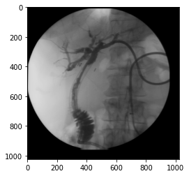
Accessing the image array
img_xray = dicom_ds.pixel_array

img_xray.shape
(1024, 1024)
plt.imshow(img_xray, cmap="gray", vmin=0, vmax=255)

plt.colorbar()
<matplotlib.colorbar.Colorbar at 0x7f617d2a9da0>
Another example
Example available in documentation.
# authors : Guillaume Lemaitre <g.lemaitre58@gmail.com>
# license : MIT
import matplotlib.pyplot as plt
from pydicom import dcmread
from pydicom.data import get_testdata_file
fpath = get_testdata_file('CT_small.dcm')
ds = dcmread(fpath)
# Normal mode:
print()
print(f"File path........: {fpath}")
print(f"SOP Class........: {ds.SOPClassUID} ({ds.SOPClassUID.name})")
print()
pat_name = ds.PatientName
display_name = pat_name.family_name + ", " + pat_name.given_name
print(f"Patient's Name...: {display_name}")
print(f"Patient ID.......: {ds.PatientID}")
print(f"Modality.........: {ds.Modality}")
print(f"Study Date.......: {ds.StudyDate}")
print(f"Image size.......: {ds.Rows} x {ds.Columns}")
print(f"Pixel Spacing....: {ds.PixelSpacing}")
# use .get() if not sure the item exists, and want a default value if missing
print(f"Slice location...: {ds.get('SliceLocation', '(missing)')}")
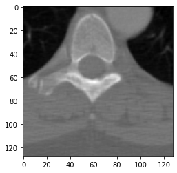
# plot the image using matplotlib
plt.imshow(ds.pixel_array, cmap=plt.cm.gray)
plt.show()
File path........: /usr/local/lib/python3.6/dist-packages/pydicom/data/test_files/CT_small.dcm
SOP Class........: 1.2.840.10008.5.1.4.1.1.2 (CT Image Storage)

Patient's Name...: CompressedSamples, CT1
Patient ID.......: 1CT1
Modality.........: CT
Study Date.......: 20040119
Image size.......: 128 x 128
Pixel Spacing....: [0.661468, 0.661468]
Slice location...: -77.2040634155
ImageIO
Load files
im = imageio.imread("0015.DCM")

im.shape
(1024, 1024)
Metadata
im.meta
Dict([('TransferSyntaxUID', '1.2.840.10008.1.2.1'),
      ('SOPClassUID', '1.2.840.10008.5.1.4.1.1.12.2'),
      ('SOPInstanceUID',
       '1.2.840.113619.2.15.1008000062035011254.825190719.0.31.2.1'),
      ('StudyDate', '19960308'),
      ('SeriesDate', '19960308'),
      ('AcquisitionDate', '19960308'),
      ('ContentDate', '19960308'),
      ('StudyTime', ''),
      ('AcquisitionTime', '105650'),
      ('ContentTime', '105650'),
      ('Modality', 'RF'),
      ('Manufacturer', 'GE MEDICAL SYSTEMS'),
      ('StudyDescription', '5'),
      ('SeriesDescription', ''),
      ('PatientName', 'Rubo DEMO'),
      ('PatientID', '10-55-87'),
      ('PatientBirthDate', ''),
      ('PatientSex', 'F '),
      ('PatientAge', ''),
      ('StudyInstanceUID',
       '1.2.840.113619.2.15.1008000062035011254.825190719.2.31'),
      ('SeriesInstanceUID',
       '1.2.840.113619.2.15.1008000062035011254.825190719.1.31'),
      ('SeriesNumber', 1),
      ('InstanceNumber', 2),
      ('PatientOrientation', ''),
      ('SamplesPerPixel', 1),
      ('Rows', 1024),
      ('Columns', 1024),
      ('BitsAllocated', 8),
      ('BitsStored', 8),
      ('HighBit', 7),
      ('PixelRepresentation', 0),
      ('PixelData',
       b'Data converted to numpy array, raw data removed to preserve memory'),
      ('shape', (1024, 1024)),
      ('sampling', (1.0, 1.0))])
im.meta["BitsAllocated"]
8
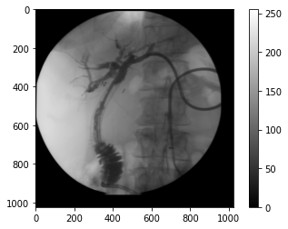
Accessing the image array
im
Array([[46, 49, 46, ...,  0,  0,  0],
       [ 0,  0,  0, ...,  0,  0,  0],
       [ 0,  0,  0, ...,  0,  0,  0],
       ...,
       [ 0,  0,  0, ...,  0,  0,  0],
       [ 0,  0,  0, ...,  0,  0,  0],
       [ 0,  0,  0, ...,  0,  0,  0]], dtype=uint8)
plt.imshow(im, cmap="gray", vmin=0, vmax=255)
<matplotlib.image.AxesImage at 0x7f617cd37940>